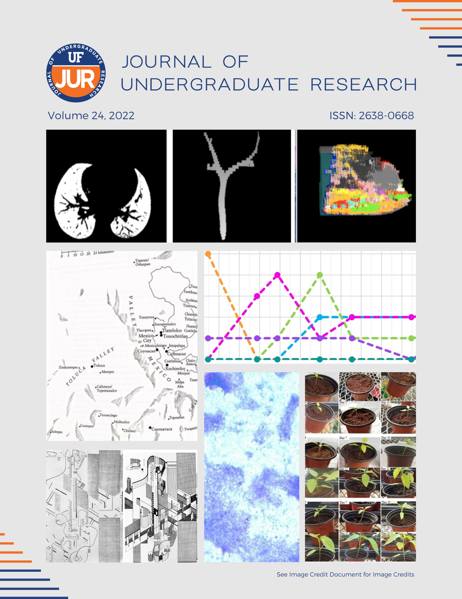
Assessing Lung Vasculature Development and Application to Early Preterm Gestation Patients
DOI:
https://doi.org/10.32473/ufjur.24.130800Keywords:
premature infant, lung vessel development, x-ray computed tomography, vessel segmentationAbstract
Individuals who were born prematurely undergo many difficulties from the time they are born to their adult lives. Obstructive lung diseases are a common occurrence in patients who were born prematurely. Unfortunately, the development of lungs in premature infants is an area in the science world that has not been fully studied. Therefore, this study’s objective is to determine trends in lung vessel growth as a patient advances in age, and discover quantitative measures that provide clinical insight regarding improving the quality of life of premature infants. This is a retrospective study where x-ray computed tomography (CT) scans were analyzed by using in-house software built upon the NIH ImageJ platform. For each scan, the lung volume was automatically identified and the pulmonary vessel trees extracted and characterized to quantify the total number of vessels in the left hemi-lung. The radius and length of each vessel tree were also recorded. CT scans of 16 pediatric patients were analyzed (7 prematurely born and 9 full-term). The vessel growth of full-term patients trended to increase as a patient aged, while vessel growth for premature patients decreased with age. The data also indicated a significantly larger difference in the number of vessel branches over time between female preterm and full term subjects. Limitations such as variations in image quality, children’s age, and changes in CT technology over time are being addressed to improve confidence in the results. Future works will include analysis on a larger data set and novel approaches to automatic vessel extraction from chest CTs.
Downloads
Published
Issue
Section
License
Copyright (c) 2022 Randi Dias, Aren Singh Saini, Walter O'Dell

This work is licensed under a Creative Commons Attribution-NonCommercial 4.0 International License.
Some journals stipulate that submitted articles cannot be under consideration for publication or published in another journal. The student-author and mentor have the option of determining which journal the paper will be submitted to first. UF JUR accepts papers that have been published in other journals or might be published in the future. It is the responsibility of the student-author and mentor to determine whether another journal will accept a paper that has been published in UF JUR.

