
Manual strangulation of a stray cat:
linking pathologic findings with the crime
Abstract: Veterinary forensic pathology plays a crucial role in animal death investigations. Recent years have seen increased attention given to this field in Taiwan. Here we report a grave crime that aroused public indignation in Taiwan, when in December 2015, a missing free-roaming cat was allegedly strangled by a suspect, according to witnesses. Surveillance video footage of the cruel incident surfaced that showed the cat, without visible signs of struggle, strangled by both hands of the suspect and kicked in the abdomen’s left side. The body was stored in a travel bag when found in the suspect’s motorcycle trunk three days after the cat had been missing. The radiographs revealed a suspected luxation of cervical vertebrae 6 and 7 (C6–C7). At forensic necropsy and histopathology, neck compression with subcutaneous hemorrhage at the left submandibular area, bruising of the neck, tears in the wall of the left external jugular vein, and pulmonary lesions were identified, all consistent with asphyxiation. Hemorrhages of the epidural space of the C7 vertebra, the spinal nerve at the level of C7, and surrounding soft tissues were noted. The pathological findings were in line with the suspect’s confession, the witnesses’ statements, and the video footage. The process and outcome of this case reflected society’s growing awareness of animal welfare and the increased attention that the authorities are giving crimes against animals in Taiwan, and the link between pathologic findings and the crime.
Keywords: veterinary forensic sciences, pathology, animal cruelty, strangulation, asphyxiation, domestic cat
The advancement of animal welfare is a worldwide trend, and this has brought with it the development of veterinary forensic sciences. Veterinary forensic pathology, an emerging discipline, plays a crucial role in animal death investigations. Each death tells its own story influenced by numerous extrinsic and intrinsic factors, and the presence of injuries varies from case to case. In forensic cases, the reference data should be applied with the awareness that individual exceptions may exist (Dolinak et al. 2005). Therefore, searching for sufficient supporting evidence and avoiding arbitrary opinions becomes the most challenging task in forensic pathology, especially in murder cases. In veterinary forensic pathology, variations among species and breeds often make the challenges more complicated. Limited research and case studies on selected types of injury further add to this situation. Furthermore, a lack of background information or evidence-based investigation often increases the difficulty of some cases, especially when the lesions might be subtle, such as those seen as a result of strangulation.
Strangulation lesions that have been reported in animal cases include the ligature mark, abrasions or muscle contusions, soft tissue hemorrhage of the neck, laryngeal-hyoid fractures, conjunctival petechiae, pulmonary edema, congestion or hemorrhage, atelectasis, emphysema, and lesions of acute respiratory distress syndrome; however, none of these lesions are pathognomonic (Munro and Munro 2008; Merck 2012; Bradley 2016; McEwen 2016, 2018). The critical objective of forensic necropsy for suspected cases of strangulation lies in finding sufficient evidence of compression to support the suspicion. Actually, in many cases, especially when the police authorities hold negative attitudes because of scanty evidence, the necropsy findings may initiate a subsequent investigation.
Strangulations are not uncommon in animals and may occur under various circumstances (Munro and Thrusfield 2001a, 2001b; Merck 2012; McEwen 2016, 2018). According to the authors’ experience, strangulation accounted for 4 of 100 forensic necropsy cases of alleged animal cruelty during 2011–2016 (WHH and CHL, unpublished data). However, according to our previous study with a real-time reporting system for causes of death in canine and feline animal cruelty cases, we found that there may be underreporting of trauma-related deaths in Taiwan (Huang et al. 2018).
Here we report a grave crime that aroused public indignation in Taiwan in 2015. In this sensational case, the offender confessed to catching and manually strangling the victim. The pathological findings supported strangulation as the cause of death. The process and outcome of the case reflect the current situation of animal welfare and veterinary forensics in Taiwan.
Case Report
In 2015, the disappearance of a male castrated crossbred free-roaming cat, named “Big Orange,” drew public attention. According to a witness, a suspicious man wearing black gloves was interacting with the cat on the afternoon of December 28. Later that afternoon, another woman witnessed the same man attempt to strangle a different street cat, which luckily escaped when the witness intervened to stop the suspect. Netizens launched a cyber search for the suspect, who was later discovered to be a senior university student from China. The police soon apprehended the suspect.
During detention, the suspect denied committing the crime. Later, surveillance video footage of an underground parking lot emerged. In the video, the suspect entered the parking lot, holding the cat by the forelimbs. The cat showed no visible signs of struggle but was still alive, as evident by a whipping movement of the cat’s tail. The suspect then manually strangled the cat for a few seconds while holding it on the ground of the parking lot; the cat became motionless. The suspect then forcibly kicked the left side of the cat’s abdomen; the cat remained motionless. The suspect left the parking lot and re-entered about 80 minutes later to put the cat, motionless, in a black travel bag. The suspect left the parking lot carrying the bag.
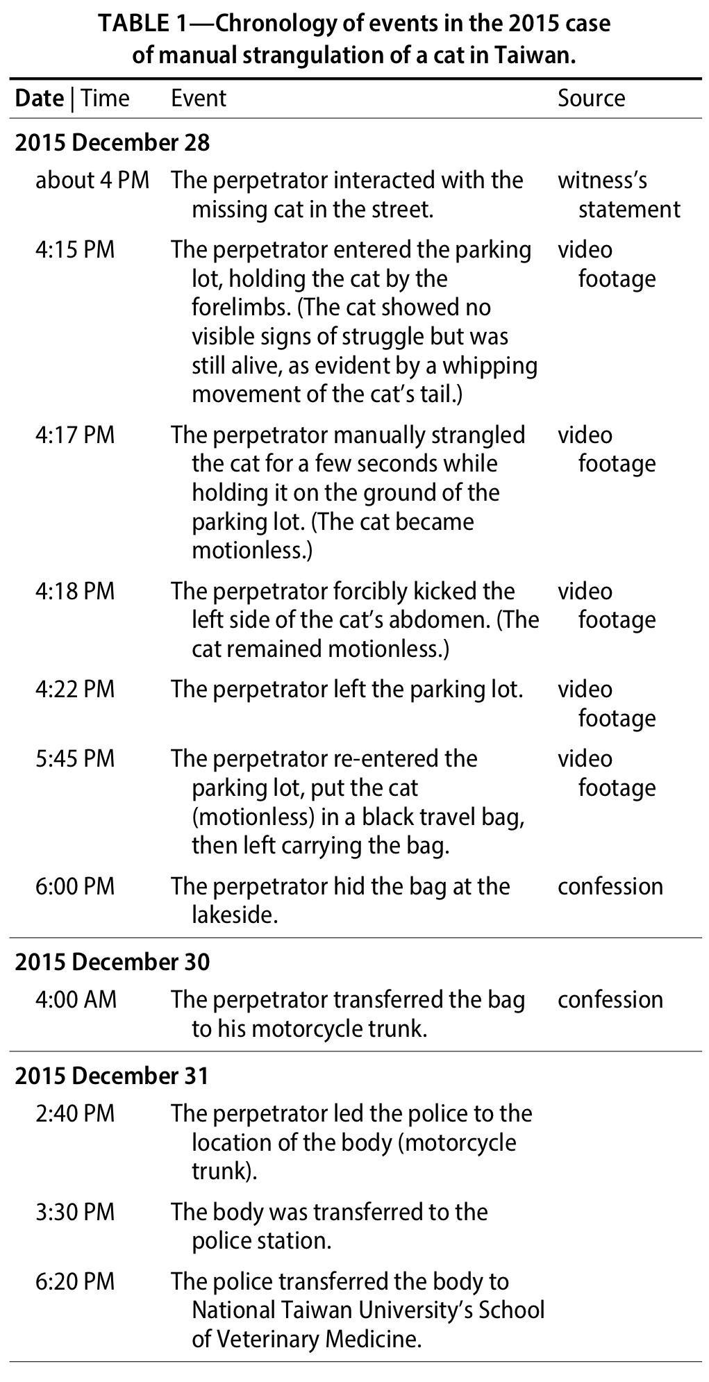
After the video footage was discovered, the suspect confessed that he had manually strangled the cat and that the body was first dumped near a lake then later transferred to his motorcycle trunk. The body was found on 31 December 2015, three days after the cat went missing. The body, still in the travel bag, was submitted to National Taiwan University’s School of Veterinary Medicine for forensic necropsy on the evening of 31 December 2015. Table 1 summarizes the chronology of significant events in the case, including the suspect’s actions and the transportation of the body.
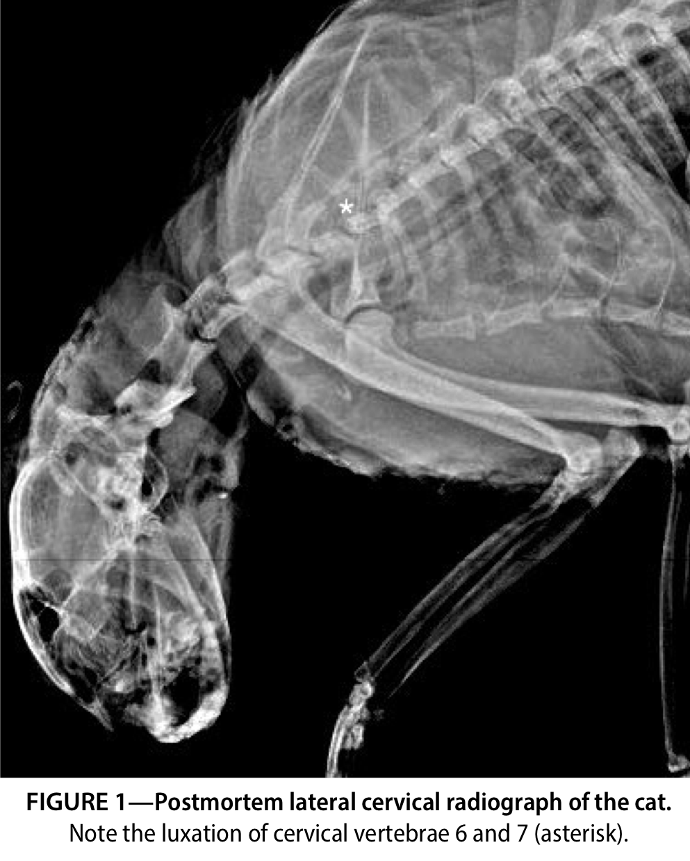
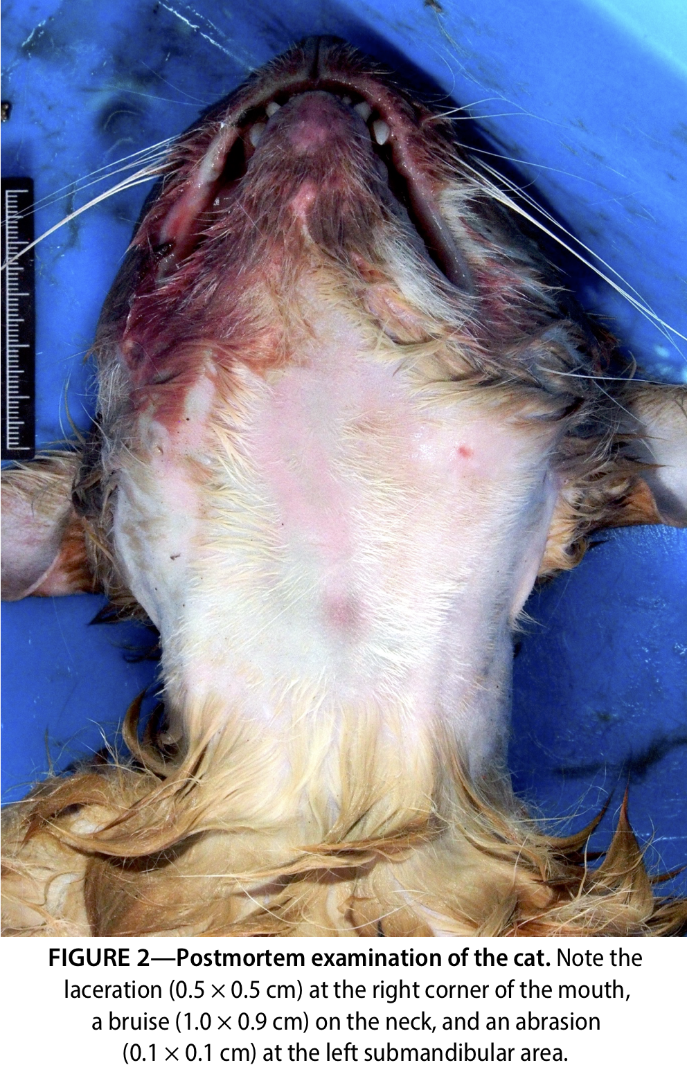
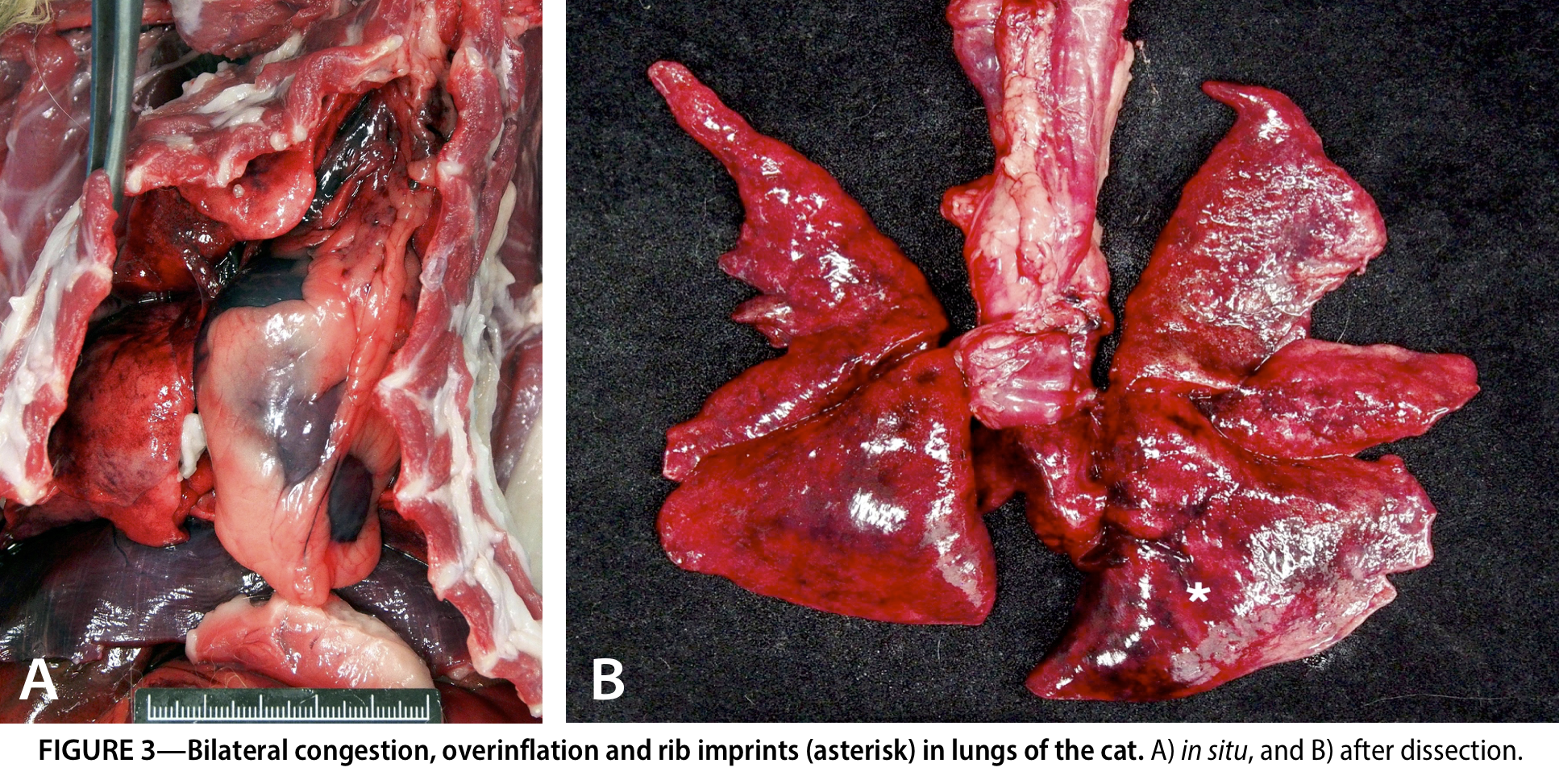
Postmortem radiography of the carcass showed luxation at C6–C7 vertebrae ( Figure 1 ). Forensic necropsy with layered neck dissection was performed. The fur was soaked and soiled with plant material and textile fibers from the travel bag. The carcass showed neither rigor mortis nor noticeable livor mortis. No petechial hemorrhages were observed at the conjunctiva, oral mucosa, or scalp. After the haircoat was clipped, a 0.5 × 0.5 cm laceration was observed at the right corner of the mouth. A 1.0 × 0.9 cm bruise on the neck and a 0.1 × 0.1 cm abrasion were noted at the left submandibular area ( Figure 2 ). Subcutaneous hemorrhage was noted around the left submandibular area, and the left submandibular lymph node was dark red. The laryngeal cartilages and hyoid apparatus were intact. The cervical muscles, internal jugular veins, and carotid arteries presented no significant lesions. Bilaterally the lungs were congested, overinflated, and had rib imprints ( Figure 3 ). Notable hemorrhage was seen in the epidural space and surrounding soft tissues at the level of the C7 vertebra, the subcutaneous tissues of the left flank, and the mediastinal soft tissues.
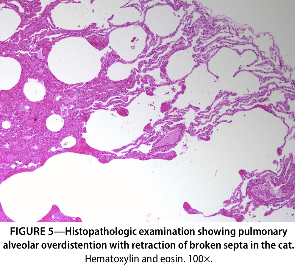
Histologically, discontinuities within the wall (between the tunica intima and tunica media) of the left external jugular vein suggested tears ( Figure 4 ). Marked parenchymal crush, with severe congestion and minimal hemorrhage, was present at the left submandibular lymph node. Alveolar overdistention, with retraction of broken septa ( Figure 5 ), and alveolar hemorrhage and severe pulmonary edema were noted. Moderate hemorrhage, without identifiable lesions of the spinal cord, was present at the C7 spinal nerve and surrounding soft tissues. Neuronal satellitosis was diffusely increased in the cerebral cortex.

The forensic necropsy findings were most consistent with manual strangulation as the cause of death, by the mechanism of asphyxiation in the manner of non-accidental injury, and were compatible with the suspect’s confession, the witnesses’ statement and the video footage. The university suspended the perpetrator and ordered him to have a mental health evaluation. In June 2016, before the case went to trial, the perpetrator was suspected of having killed a second free-roaming cat. Again, the violent actions of the cat killer aroused public indignation. The perpetrator admitted to both killings and dumping the second cat in a river. The body of the second cat has not been recovered. In October 2016, the perpetrator was sentenced to 10 months in prison and fined NT$350,000 for the killing of two cats.
Discussion
In this article, we present a highly sensational 2015 case of a manually strangulated cat in which the forensic necropsy findings provided explicit evidence of the offender’s violent crime. The case influenced the amendment of animal protection laws. The process and outcome of the case reflect the current situation of animal welfare and veterinary forensics in Taiwan.
Aside from determining the cause, mechanism and manner of death, another mission of forensic pathologists is to answer legal questions raised by the authorities, and one should prepare for any questions that may be asked in the future (Dolinak et al. 2005). In the present case, despite proof of manual strangulation being evident, several questions are worth discussing.
First, what happened to Big Orange? Big Orange was last seen alive in the video footage of the parking lot after the offender had been observed interacting with the cat on the street about fifteen minutes prior. Based on the necropsy with histopathology, proof of neck compression included the subcutaneous hemorrhage at the left submandibular area, the bruise on the neck, and tears in the wall of the left external jugular vein. These may be associated with the perpetrator’s actions of strangling the cat that was seen on the video footage. Hemorrhages in the left flank could also be associated with the perpetrator’s action of kicking the cat, which was caught on video, and this vital reaction suggests that the cat was alive when it was kicked.
This finding leads to another question: was the death caused by manual strangulation or by other causes, such as drowning or environmental asphyxia due to oxygen depletion in the enclosed environment of the travel bag. Since the cat was put into the travel bag without other observable attacks after the kick, the victim may have died within the bag. It has been reported that dogs and cats could remain conscious for some time after being strangled because of the anatomic variations in blood supply to the brain compared to humans (McEwen 2018). Besides, strangulation rarely seems to cause complete cerebral ischemia in dogs and cats (McEwen 2018).
According to previous studies, the presence of neuronal satellitosis in the cerebral cortex may be associated with the ischemic conditions (Oehmichen et al. 2006, Olczak et al. 2018). In this case, being placed into the travel bag could have created an oxygen-depriving environment leading to the development of satellitosis in the cerebral cortex. It is also well known that the overinflation of lungs with rib markings and the alveolar overdistention with the retraction of broken septa have been observed in cases of drowning in both humans and animals. The fact that the cat was initially left at the lakeside, along with the necropsy findings (soaking fur and pulmonary lesions), raises the suspicion of drowning. However, it has been reported that the overinflated lungs with rib markings and alveolar over-distention could also be observed in animal cases of strangulation (Munro and Munro 2008). Although the histological features found in the lungs were in line with a previous case report on the manual strangulation of a cat (Bradley 2016), it is not possible to differentiate the causes of asphyxia by the pulmonary lesions alone (Delmonte and Capelozzi 2001). Besides, according to the information the authors obtained, it was unclear whether the travel bag was entirely closed or whether the cat was immersed in the water when it was alive. However, it was known that the body was at the lakeside for more than 24 hours and was soaking wet when found. Therefore, it was still inconclusive whether the death was caused by drowning or environmental asphyxia, and we cannot rule out the possibility that the cat might have died from multiple causes of asphyxia. The cat may likely have suffered longer than what we expected.
Another interesting question is why the cat showed no visible signs of struggle on the video footage. A partial explanation for this may be due to the injuries at the level of C7 vertebra, which might have caused paralysis, decreased spinal reflexes and decreased muscle tone of the forelimbs, and significant pain (de Lahunta et al. 2014). With these findings, it is possible that the perpetrator had already strangled and severely injured the cat before entering the parking lot.
The link between animal abuse and interpersonal violence has drawn more interest and attention recently (Ascione et al. 2007; Lockwood and Arkow 2016; Rogers and Stern 2017). A checklist has been established to evaluate the significance of an individual’s involvement in a particular act of animal cruelty as an indicator of the dangerousness of the participation of future acts of violence against humans (Lockwood and Arkow 2016). Our perpetrator meets multiple criteria in this checklist, which implies that he may engage in interpersonal violence in the future.
The process and outcome of this case demonstrated several advancements in animal welfare and veterinary forensics. First, this may be the most severe penalty for animal cruelty in recent years in Taiwan. Second, concerned citizens and government authorities participated in the case and reacted efficiently, compared to previous crimes against animals. Third, the public showed a higher level of attention and compassion for the animal victim. Finally, the Taiwanese system for animal death investigation vastly improved as a result of this case. These improvements reflect society’s growing awareness of animal welfare and the increased attention that the authorities are giving crimes against animals in Taiwan.
Acknowledgments
The article was funded by Taipei City Animal Protection Office under grant no. 104 and by Ministry of Science and Technology, Taiwan, R.O.C. under grant no. MOST103-2313-B-002-041-MY3. The authors gratefully acknowledge Aurora Animal Hospital for sponsoring the radiography and Institute of Forensic Medicine, Ministry of Justice, Taiwan (Republic of China) for assisting in pathologic diagnosis.
Disclosures
None of the authors of this paper have a financial or personal relationship with other people or organizations that could inappropriately influence or bias the content of the article.
References
- Ascione FR, Weber CV, Thompson TM, Heath J, Maruyama M, Hayashi K. 2007. Battered pets and domestic violence: animal abuse reported by women experiencing intimate violence and by nonabused women. Violence against women. 13(4):354–73. doi:10.1177/1077801207299201.
- Bradley N. 2016. Asphyxia: the unusual tale of two cases. North American Veterinary Community conference; 2016 Jan 16–20; Orlando. NAVC. p. 353–5.
- de Lahunta A, Glass EN, Kent M. 2014 . Veterinary neuroanatomy and clinical neurology. Saunders, UK.
- Delmonte C, Capelozzi VL. 2001. Morphologic determinants of asphyxia in lungs: a semiquantitative study in forensic autopsies. Am J Forensic Med Pathol . 22(2):139–49. doi:10.1097/00000433-200106000-00006 .
- Dolinak D, Matshes E, Lew EO. 2005. Forensic pathology: principles and practice. Academic Press, Netherlands.
- Huang WH, Liao AT, Chu PY, Zhai SH, Yen IF, Liu CH. 2018. Trauma-related deaths of domesticated dogs and cats in Taiwan. Taiwan Vet J . 44(01):15–26. doi:10.1142/S1682648517500111 .
- Lockwood R, Arkow P. 2016. Animal abuse and interpersonal violence the cruelty connection and its implications for veterinary pathology. Vet Pathol. 53(5):910–8. doi:10.1177/0300985815626575 .
- McEwen BJ. 2016. Nondrowning asphyxia in veterinary forensic pathology: suffocation, strangulation, and mechanical asphyxia. Vet Pathol. 53(5):1037–48. doi:10.1177/0300985816643370 .
- McEwen BJ. 2018. Strangulation, suffocation, and asphyxia. In: Brooks JW, editor. Veterinary forensic pathology. Vol. 1. Cham (Switzerland): Springer. p. 129–48. doi:10.1007/978-3-319-67172-7_8 .
- Merck M. 2012. Veterinary forensics: animal cruelty investigations. Wiley, UK. Munro R, Munro HMC. 2008. Animal abuse and unlawful killing: forensic veterinary pathology. Saunders, Philadelphia, PA. doi:10.1016/B978-0-7020-2878-6.X5001-6 .
- Munro HM, Thrusfield MV. 2001a. 'Battered pets': features that raise suspicion of non-accidental injury. J Small Anim Pract. 42(5):218–26. doi:10.1111/j.1748-5827.2001.tb02024.x .
- Munro HM, Thrusfield MV. 2001b. 'Battered pets': non-accidental physical injuries found in dogs and cats. J Small Anim Pract. 42(6):279–90. doi:10.1111/j.1748-5827.2001.tb02041.x .
- Oehmichen M, Auer RN, König HG. 2006. Forensic neuropathology and associated neurology. Springer, Berlin, Heidelberg. doi:10.1007/3-540-28995-X .
- Olczak MD, Chutorański D, Kwiatkowska M, Samojłowicz D, Tarka S, Wierzba-Bobrowicz T. 2018. Bystin (BYSL) as a possible marker of severe hypoxic-ischemic changes in neuropathological examination of forensic cases. Forensic Sci Med Pathol. 14(1):26–30. doi:10.1007/s12024-017-9942-x .
- Rogers E, Stern AW. 2017. Veterinary forensics: investigation, evidence collection, and expert testimony. CRC Press, Boca Raton, FL. doi:10.4324/9781315153421 .